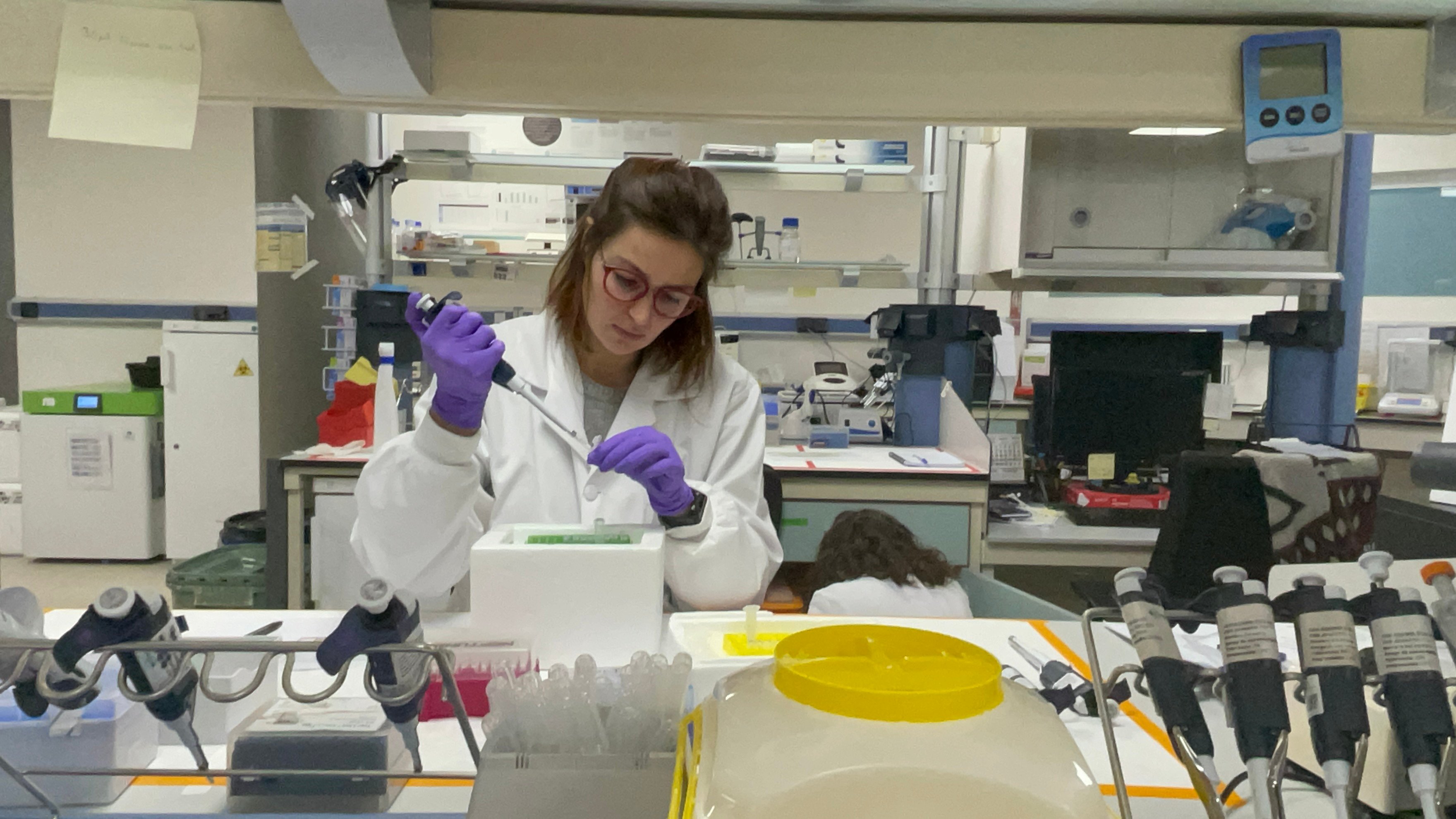
A wide variety of substances like chemicals, pollutants, or drugs can damage the nervous system, causing neurotoxicity and leading to neurological disorders. Thus, there is an urgent need to test the neurotoxicity of many chemical compounds people are exposed to and which effects on nervous systems are unknown.
In this sense, the standard methods for neurotoxicity testing using rodent models cannot keep pace with the increasing number of substances in continuous development that need to be tested, as they are costly, also require significant space, and entail ethical concerns. To overcome these limitations, alternative models in neurotoxicity have been developed, including computational, in vitro, and in vivo models.
Although computational and in vitro models offer several advantages, such as speed, reduced cost, the complete abolition of animal use, and the ability to screen a large number of substances, they cannot fully replicate the complex interactions that occur in vivo. Several organisms are being used as alternative in vivo models for neurotoxicity, from invertebrates such as Drosophila melanogaster or nematode, to vertebrates like zebrafish.
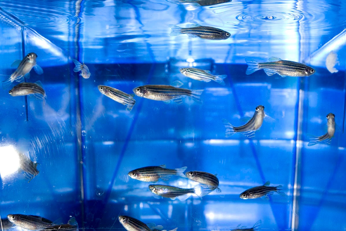
The zebrafish has become a powerful tool to overcome the limitation of other in vivo models and are increasingly being used as a model organism for neurotoxicity.
At Biobide, we have developed a rapid and non-invasive Neurotoxicity Assay using zebrafish with high sensitivity and 100% specificity, that is suitable for use in early neuroactive drug screenings.
Neurotoxicity
Neurotoxicity is the capacity of substances or physical agents to damage the nervous system (brain, spinal cord, and peripheral nerves), causing functional or structural alterations that may lead to various neurological disorders. The symptoms of neurotoxicity can vary, depending on the nature and extent of the damage, and may include changes in behavior and cognitive impairment. Thus, preventing neurotoxicity requires the identification of harmful substances, to reduce exposure to them, as well as to define appropriate safety precautions.
Chemicals that are commonly associated with neurotoxicity include heavy metals and organic solvents. Some drugs for example antipsychotics, antiepileptics, and those used for chemotherapy are also associated with neurotoxicity.
Drugs can cause neurotoxicity by several mechanisms, some cause direct damage to the nervous system cells and others cause indirect damage by the induction of harmful physiological events such as oxidative stress, inflammation, or disruption of normal cellular processes. Some of the common effects of neurotoxicity produced by drugs include cognitive impairment, behavioral changes (aggression, depression, anxiety, and mood swings), movement disorders, peripheral neuropathy, and seizures.
Due to the complexity of the nervous system and the outcome produced by neurotoxicity, it is necessary to use in vivo models that can recapitulate the damage observed in the human nervous system. For that, the model organism must replicate the brain’s physiological and structural features, the presence of immune and blood-brain barrier interactions, and the ability to present a complex behavior.
Evidently, the best organisms to test neurotoxicity are mammals because of their high genetic homology with humans which produces very similar nervous systems. However, the use of animals with complex nervous systems has ethical dilemmas, is time-consuming, and highly priced. As a result, there is an unmet need for fast, sensitive, and cost-effective New Alternative Models (NAMs) to predict neurotoxicity.

Alternative models for neurotoxicity
The selection of an appropriate model organism is particularly important in neurotoxicity studies and different alternative models have been developed to overcome the limitation mentioned.
Computational models, such as molecular docking, quantitative structure-activity relationships, and artificial neural networks can predict neurotoxicity based on the chemical structure and physicochemical properties of a substance. These models offer several advantages, such as speed, cost-effectiveness, the ability to screen a large number of substances, and the avoidance of the usage of living organisms. Nevertheless, they have limitations, such as the need for high-quality input data, the need for validation and optimization, and the inability to fully replicate the complex interactions that occur in vivo.
In vitro and in vivo models have been extensively used to conduct toxicity tests. Cell lines are the most frequently used models for in vitro systems because they are inexpensive and allow large-scale screening. Human-derived cell-based models use induced pluripotent stem cells differentiated into neurons and glia to mimic the complex structure and function of the human brain. Organotypic slice cultures can reproduce the complex three-dimensional structure of the brain, while microfluidic systems can mimic the microenvironment of the brain, including the blood-brain barrier, immune cells, and extracellular matrix. These models offer several advantages, including being low cost, ease of use, and the ability to screen a large number of substances rapidly. However, they have limitations such as replicating the complex structures of the brain, lack the ability to study systemic effects, and are not capable of representing behavioral outputs.
Therefore, in vivo models nowadays are irreplaceable for assessing neurotoxicity. Thus, there is a wide range of organisms being used as alternative in vivo models for neurotoxicity including invertebrates like Drosophila melanogaster or nematode and vertebrates like zebrafish. Drosophila has a simple nervous system and can be genetically manipulated, making them useful for studying the underlying mechanisms of neurotoxicity. Nevertheless, since invertebrates have no complex neural system structure such as myelin sheath, the research on neural activity is limited.
On the other hand, zebrafish are increasingly being used as model organisms for neurotoxicity studies. They possess rapid development in the embryonic phase, high fecundity, highly transparent embryos, and over 80% of zebrafish genes are homologous to humans. Moreover, their nervous system arrangement and the process and specific mechanisms of neurogenesis are similar to other vertebrates, including humans, also they have a Blood Brain Barrier which is of added value when conducting assays in the field of neurotoxicity and overall becoming an excellent animal model for neurotoxicology research as recapitulate with high fidelity human nervous system.
Zebrafish as model for neurotoxicity
Zebrafish has become a powerful tool to overcome the limitations of other models in neurotoxicity testing. The small size allows placing them in standard microplates to perform High Content screenings. Importantly, the results of the zebrafish screens show a good correlation with mammalian models of toxicology supporting the utility of this model. Besides, the use of zebrafish larvae is in accordance with the 3R principles and is considered a New Alternative Method (NAM) because they can be used in the embryonic stage as they present fast organogenesis and can replace mammals in many experiments.
Comparative neurogenetic and neuroanatomical analyses reveal high degrees of conservation between the nervous systems of zebrafish and mammals. Mammalian brain subdivisions, such as the telencephalon, diencephalon, mesencephalon, and rhombencephalon have zebrafish counterparts. Additionally, zebrafish and mammals share developmental processes, such as neurogenesis, axon guidance, and relevant genetic signaling.

Several approaches using zebrafish in neurotoxicity screening have been developed. Neurotoxicity endpoints such as changes in gene expression, neural morphogenesis, and neurobehavioral profiling are used to assess the effects of substances on the nervous system. Alterations in the mobility pattern of larvae induced by known drugs have been observed to strongly replicates what is observed in humans, supporting the use of zebrafish as a predictive model of neuroactivity in humans. Behavioral profiling in zebrafish reveals relationships between drugs and their targets and demonstrates a conserved vertebrate neuropharmacology.
The central nervous system in zebrafish is mostly developed within 3 days of fertilization (dpf), making them ideal for rapid neurotoxicity tests. At this stage they also show spontaneous swimming behavior, allowing the automated assessment of locomotor activity under different conditions. Subsequently, at 5 dpf, embryos present a quite developed nervous system being able to respond to several external factors such as light changes, noises, or vibrations.
A common behavioral assay for zebrafish larvae activity analysis consists of tracking larvae movement within well plates of different sizes while modifying environmental conditions, such as alternating light and dark phases, zebrafish are a diurnal species and have a fully regulated circadian system by 6 dpf due to the melatonin synthesis, stablishing them as an ideal model for Behavior Alteration Assays.
Therefore, the use of zebrafish in the larval stage, before 6 dpf, offers the possibility of testing neurotoxicity in a vertebrate organism without the ethical concern associated with the use of these animals.
Neurotox Assay
The morphological and behavioral changes in zebrafish are direct macroscopic evidence of the neurotoxic effects of compound exposure. Biobide has set up a High Content assay to evaluate general behavioral alterations in 5 dpf zebrafish larvae, based on responses to dark-light changes. The assay was validated with a total of 28 drugs with different human therapeutic indications such as fluoxetine, MPTP, dopamine, and norepinephrine, obtaining a sensitivity of 95% and a specificity of 100% compared with their effect in humans.
This assay measures several locomotor parameters, based on the speed and swimming pattern, in response to the presence or absence of light; allowing a complete evaluation of behavioral alterations related to neurotoxicity. Furthermore, the test is performed on 96 well plates, allowing a fast, scalable, and cost-effective assay.
Biobide’s Neurotox Assay is an alternative, rapid, cost-effective, and non-invasive assay with high sensitivity and 100% specificity that is suitable for use in early neuroactive drug screenings.
Conclusion
The evaluation of the toxic effects on the nervous systems caused by new chemical compounds, also the harmful effects by already commercial products, whose effect have not been properly analyzed is a first-order problem that requires the development of reliable, fast, highly scalable, and competitive testing methods. In addition, new ethical concerns linked to the use of animals for laboratory tests drive the development of New Alternative Methods (NAMs).
Due to the complexity of the human nervous system, zebrafish embryos have been postulated as a useful alternative method for neurotoxicity testing because of the similarity between their nervous system compared to humans, the small size they present, their easy and affordable raising, the competitive cost-effectiveness, and the less ethical concerns they present.
In this sense, Biobide has developed a High-Content neurotoxicity assay that enables a fast, cost-efficient, and complex behavioral analysis in response to changes in light intensity to accelerate and promote the evaluation of the neurotoxicity of the chemical compounds that people are exposed to.
Sources
- https://pubmed.ncbi.nlm.nih.gov/30521027/
- https://biobide.com/portfolio/neurotox-assay
- https://pubmed.ncbi.nlm.nih.gov/36581008/
- https://pubmed.ncbi.nlm.nih.gov/35870347/
- https://zantiks.com/protocols/light-dark-locomotion-test-larval-zebrafish


