
Transgenic animal models are an important tool for the discovery of human disease-related mutations as well as the drugs that treat them. Genetic disorders found in humans can often be mimicked in other living organisms and used in large scale throughput screenings to identify possible therapeutic compounds. Transgenic zebrafish are one of those in vivo models that have become popular for researchers, including rare disease research.
In this article, we’ll explore transgenic fluorescent zebrafish lines in particular, and how they can be used in Drug Discovery and development.

Advantages of Fluorescent Zebrafish in Drug Screening
As a teleost class of fish species, zebrafish don’t appear to have much in common with humans. Under the ‘hood’, however, there is sufficient homology as vertebrates in biochemical pathways and organ structures between zebrafish and humans. Astoundingly, despite our obvious differences, zebrafish have orthologs in 80% of human disease related genes. Also, the zebrafish genome has been totally sequenced. Zebrafish genes are therefore easily manipulable, and over 14,000 mutations have been created in zebrafish genes. This means zebrafish are exceptionally well suited to genetic disease research.

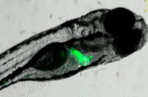
Image of a transgenic zebrafish Tg(wt1b:EGFP), expressing fluorescent protein e-GFP in kidney progenitor cells. Source
Moreover, the embryonic and larval stages of zebrafish offer even more advantages, such as external development and fertilization, embryo transparency, and rapid organogenesis (totally developed by 48-96 hours post-fertilization). The combination of these features means that researchers can even observe development at the single cell level. Zebrafish are therefore ideal for expressing fluorescent proteins (GFP, m Cherry...) to mark a concrete target or organ for image analysis.
All of these characteristics lend themselves to an easy observation by researchers, allowing them to develop Phenotypic screenings of this animal model in large scale in vivo studies.
Reverse genetics methods have allowed researchers to alter orthologous genes that are possibly related to human diseases. Fluorescent zebrafish transgenic lines are utilized in a wide variety of fields like toxicology and efficacy assays, which we will discuss below.

Application of Fluorescent Transgenic Zebrafish Lines
Due to the transparency of zebrafish embryos, it is possible to conduct an analysis using fluorescent proteins. The advantages of this model include assessing the potential toxicity of pharmacological, agrochemical, and cosmetic compounds, as well as studying the efficacy of new drugs.
Toxicology Assays:
Zebrafish with fluorescence in a target organ are well suited for studying the toxicity related to those organs through image analysis. Analysis can be done not only qualitatively but also by quantifying the fluorescence. This fluorescence may emit in multiple wavelengths depending on the protein used to generate them. One of the most used proteins is GFP, which generates a green fluorescence, and mCherry, which generates a red fluorescence.
Cardiotoxicity
Cardiotoxicity, in particular, continues to be a major threat to human health, especially concerning heart development in the early stages of life. It is also one of the main causes of withdrawal of drug candidates in preclinical stages. In terms of heart development and biology, zebrafish hearts are very similar to human ones, with the difference being that Zebrafish have only one atrium and one ventricle instead of two of each.
Zebrafish with green fluorescence in the heart allows researchers to track the heart beating and makes it possible for them to record a video of the heart function. This allows researchers to identify different heart functional alterations, such as arrhythmias or atrioventricular failures. The 1:2 arrhythmia specifically resembles the QT prolongation caused in humans, which can end in heart failure. Researchers have found that drugs producing cardiac toxicity in humans usually reproduce bradycardia or a 2:1 arrhythmia in zebrafish embryos, which can be analyzed in a High Content Screening (HCS) platform, thus increasing the efficiency of those pre-screening assays.
Hepatotoxicity
Along with cardiotoxicity, hepatotoxicity is one of the main causes of withdrawal of drugs in early Drug Discovery. Transgenic zebrafish with fluorescence in the liver allows you to visualize and identify the whole area of the liver better and identify liver cells by image analysis.
Nephrotoxicity
Nephrotoxicity is another area where Zebrafish are a key tool in drug research. Kidney disease is a serious global problem. Fluorescent Zebrafish are increasingly used to analyze kidney function due to their similarities to humans. Like the heart, their renal anatomy is very comparable to humans. They are used to screen for nephrotoxicity and therapeutic compounds for kidney diseases. Transgenic zebrafish with fluorescence in the kidney helps to visualize the organ better and detect malformations of the glomerulus or the tubules.
Efficacy Assays: Neurodegenerative and Neuromuscular Diseases, Oncology, Inflammatory and Cardiovascular diseases
Several neurodegenerative and neuromuscular zebrafish models have been created and can be used for target validation and to screen potential drugs to treat these diseases. Many of them are integrated among the rare diseases because they affect a small number of people compared to the general population.
Several transgenic fluorescent zebrafish models can be used to study neurodegenerative diseases, allowing researchers to develop in vivo screenings that enhance therapy research. These diseases include:
- Parkinson's Disease
- Alzheimer's Disease
- Epilepsy
- Huntington's disease.
- Muscular dystrophies
- Amyotrophic Lateral Sclerosis (ALS)
- Duchenne Muscular Dystrophy (DMD)
- Dravet Syndrome (DS)
- Usher Syndrome.
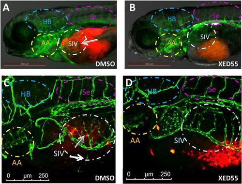
Fluorescent expression of blood vessels in transgenic zebrafish for angiogenesis research. Source
Transgenic fluorescent zebrafish are also promising models in the fight against several cancers. Angiogenesis refers to the development of new blood vessels from the existing vasculature and blocking this vessel growth is one way to fight against tumor development. Drug screening using transgenic zebrafish, which express green fluorescent proteins in blood vessels, helps to identify chemicals that impact tumor growth.
In conclusion, transgenic fluorescent zebrafish lines expressing fluorescent proteins offer unique features such as productivity, reliability, embryos transparency or rapid organ development, which are combined with modern image analysis tools to offer very solid screening models in biomedical research.