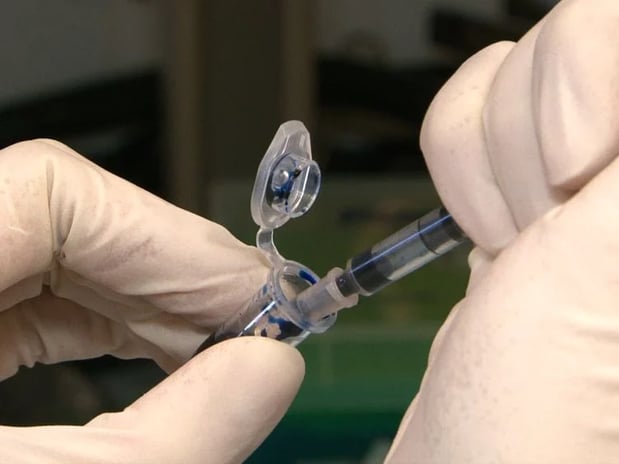
The advancement of science and technology has provided many incredible procedures, as well as discoveries that have truly helped to move humanity forward. For example, thanks to numerous scientific achievements such as MRI machines and X-ray machines we are able to detect health problems in people with great efficiency. This allows us to get patients the proper help that they need. One of those scientific advancements is none other than the use of Evans blue staining. Below we will discuss the importance of Evans blue staining and how it is greatly benefiting humanity.

History of Evans Blue Dye
Evans blue staining has a long rich history in the scientific and medical field. One of the initial staining applications was developed by Herbert Mclean Evans in 19141. Evans blue staining has been used in biomedicine for decades for a wide variety of reasons. Evans blue dye is excellent for biomedicine research because of its high-water solubility, as well as its unique ability to tightly constrain serum albumin1. In addition, Evans blue staining is widely known for its medical applications such as detection of lymph nodes, estimation of blood volume and localization of tumor lesions.
Evans Blue Dye
What is exactly Evans blue staining and how is it used? Evans blue staining or Evans blue dye is a highly extended scientific stain with medical applications in histology as well as in fluorescent microscopy2. Evans blue staining is most commonly used to check the feasibility and capability of cells2. In addition, another significant application of this incredible medical application is the ability to study blood-brain barrier permeability2. Additional scientific and medical applications of Evans blue staining include the highlighting of particular pituitary cell models, specific staining of neutrophil plasma membranes, and fluorescent microscopy as a method to reduce undesirable autofluorescence2.
Furthermore, blood vessels play a significant role in the proper functioning of the human body and its malfunctioning is related with several diseases. For this reason, researching and understanding how blood vessels work in depth is extremely critical to understand the human body3. At the same time, attempting to study blood vessels can be quite challenging, time consuming, and costly. The florescent attributes of Evans blue staining make it an indispensable tool for medical application and enables scientist to visualize and study structures such as blood vessels at a more efficient and affordable cost than traditional medical applications3.
Insertion of Evans Blue Dye
As we all know, the human body is a truly incredible construct with the ability to do many miraculous things. Did you know that there is approximately sixty-thousand miles of blood vessels in the human body3? As mentioned before, these blood vessels are essential to facilitating the functions of the body. In addition, blood vessels form an intricate, and important network which helps supply nutrients throughout the body, take out toxins, increase the immunity of the cells, and support the repair that takes place in the body after any sort of injury3. Blood vessels move blood around the human body throughout a person’s daily routine. What’s more, the blood vessels that are in the body vary in shapes and sizes3. For this reason, being able to visualize these blood vessels is critical to medical research. Evans blue staining as mentioned before, can be injected into the body, thus enabling the visualization of blood vessels and their intricate networks3.
Evans Blue Staining Coupled with The Use of Zebrafish
Zebrafish have a uniquely similar genetic structure to that of human beings. In addition, they share approximately seventy percent of genes with human beings and up to eighty-four percent of genes related to human diseases have a zebrafish counterpart. For this reason, zebrafish have become very important to medical research. Zebrafish are widely used to study vascular networks3. In addition, the transparent embryos of Zebrafish coupled with the use of Evans blue staining, as well as transgenic florescent reporters allow for the visualization of these networks3. At Biobide we know that the use of the Zebrafish model can help in a more time and cost-efficient way as opposed to other traditional methods of studying blood vessel networks within the human body.
Sources
- Hindawi, ‘Contrast Media & Molecular Imagin ’ date accessed on 6/26/2022 https://www.hindawi.com/journals/cmmi/2018/7628037/
- Milipore Sigma, ‘Evans Blue’ date accessed on 6/26/2022: https://www.sigmaaldrich.com/US/en/product/sigma/e2129
- Future Science, ‘BioTechniques’ date accessed on 6/26/2022 https://www.future-science.com/doi/10.2144/btn-2020-0152


