
Angiogenesis is a dynamic process by which blood vessels grow from pre-existing ones. The initial network is set up by vasculogenesis, the process of de novo formation of vessels, and becomes modified and grows by angiogenesis. Unlike most other organs, vasculature has a remarkable capacity to regenerate, and grow also in the adult organism, to efficiently repair damaged tissues.

Angiogenesis is fundamental in wide outspread physiological processes, including embryonic vascular development, differentiation, wound healing, and organ regeneration. This process is regulated by pro- and anti-angiogenic protein factors genes, and a disturbance of their genetic expression may lead to pathological conditions such as contribution to cancer progression, rheumatoid arthritis, or psoriasis.
Cancer is a group of complex diseases characterized by a loss of cell control mechanisms of a small group of cells caused by genetic and epigenetic alterations. These alterations transform the affected cells into uncontrolled entities able to grow limitless and disseminate through the organs, damaging them, and causing the irremediable death of the patient if it is not treated. The ability of cancer cells to grow and disseminate relies on creating new blood vessels to fulfill the additional nutrients and oxygen requirements and, therefore, angiogenesis plays a pivotal role in enabling this process.
The study of the inhibition of angiogenesis is key for developing therapeutic interventions for angiogenesis-related disorders. Angiogenesis inhibitors are a promising class of drugs that target this process, offering potential therapies for cancer and other angiogenesis-related diseases. An example is the treatment of cancer patients with the monoclonal anti-vascular endothelial growth factor-A (VEGF-A) antibody bevacizumab.
Studying these inhibitors in an efficient and cost-effective manner is crucial for advancing the understanding and developing innovative treatments. Traditional methods often involve mammalian models and are time-consuming, involve high costs, and major ethical concerns. Thus, researchers are increasingly turning to New Alternative Models (NAMs) such as zebrafish, that present unique advantages. In this article, we will explore how to study angiogenesis inhibitors using zebrafish.
Angiogenesis Inhibition
Angiogenesis is a tightly regulated process involving an intricate balance of pro- and anti-angiogenic factors. In normal conditions, pro-angiogenic factors like vascular endothelial growth factor (VEGF) stimulate the formation of new blood vessels to meet the demands of growing tissues. On the contrary, angiogenesis inhibitors act by disrupting this balance, targeting either the pro-angiogenic factors, endothelial cells, or their receptors.
The main mechanisms of angiogenesis inhibition include:
- Inhibition of VEGF signaling: VEGF is a key regulator of angiogenesis and is responsible for promoting the growth and permeability of blood vessels. Angiogenesis inhibitors can directly target VEGF or its receptors to hinder angiogenesis.
- Blocking endothelial cell proliferation: Some angiogenesis inhibitors prevent endothelial cell multiplication, which forms the inner lining of blood vessels.
- Induction of endothelial cell apoptosis: Certain inhibitors induce apoptosis (programmed cell death) in endothelial cells, leading to the regression of existing blood vessels.
- Extracellular matrix remodeling: Angiogenesis inhibitors may interfere with the breakdown of the extracellular matrix, inhibiting the migration of endothelial cells and the formation of new blood vessels.
Currently, tumor inhibition via angiogenesis targeting has been achieved by the development of drugs that belong to the classes of monoclonal antibodies (bevacizumab, cetuximab, trastuzumab, panitumumab), tyrosine kinase inhibitors (axitinib, cabonzantinib, pazopanib, regorafenib, sorafenib, sunitinib, vandetanib) and mTOR targeting compounds.

Why study angiogenesis inhibitors using zebrafish?
The zebrafish model presents many benefits and is listed by the National Institutes of Health as the third largest vertebrate model organism after mice and rats. Zebrafish embryos are emerging as an in vivo model for the study of angiogenesis in a reliable and cost-effective manner and presenting fewer ethical concerns compared with other vertebrate models, in line with the 3Rs Principle (replacement, reduction, and refinement). The anatomy of the developing vascular tree, the angiogenesis process, and the molecular mechanisms involved in vascular structure formation are strikingly similar to those found in humans and higher vertebrates. Furthermore, major molecular pathways regulating angiogenesis in mammalian systems (vascular endothelial cell growth factors, fibroblast growth factors, ephrin receptors, angiopoietins, etc.) are conserved in zebrafish.
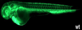
The optical transparency of the zebrafish embryo makes it easy to directly visualize and track the changes in the developing vascular system. Moreover, the development of different fluorescent transgenic reporter lines specific for vascular markers provides further tools to study the angiogenic process. Some of these transgenic lines include the Tg(fli1:egfp) with specific fluorescence in vascular endothelial cells (ECs), the Tg(lyz:egfp) with myeloid cells (MCs) specific fluorescence, and the Tg(c-myb:egfp) with hematopoietic stem cells (HSCs) specific fluorescence.
Also, zebrafish embryos are ideal to study angiogenesis and the cardiovascular system is among the first organ systems to develop and function within the developing zebrafish embryo. It consists of the heart, the blood vessels, and the lymphatic vasculature that ensures efficient circulation and thereby the supply of all tissues with oxygen and nutrients.
Angiogenesis assay usually involves treating zebrafish embryos with compounds and observing different endpoints such as the phenotypic changes in blood vessel formation, like vessel length, branching patterns, and density. Then, quantitative analysis of these phenotypic changes can be performed using specialized fluorescence microscopy or other imaging techniques.
Zebrafish can also be subjected to vascular injury, such as tail fin amputation or laser-induced injury, to study the regenerative angiogenic response. Angiogenesis inhibitors can be administered to assess their impact on the healing process and blood vessel regeneration. Additionally, analysis of angiogenesis-related gene expression can also be performed using quantitative PCR to determine the expression change of angiogenesis markers affected by the angiogenesis inhibitors.
Biobide’s Angiogenesis Inhibition Assay
Biobide has developed a High Content Screening (HCS) assay method to detect the capability of a compound to inhibit angiogenesis.
Angiogenic vessels are simply monitored, thus, making them suitable for the identification of angiogenesis inhibitors. Employing a transgenic zebrafish line with fluorescence in the blood vessels the visualization and analysis of the intersegmental vessels can be done directly even quantifying the number of vessels and their branching. After the treatment of embryos for 24 hours, fluorescent images are recorded in the microscope and fluorescent area, total number of intersegmental vessels and their number are quantified.
Biobide’s angiogenesis assay has been validated with many reference compounds with angiogenic inhibition capacity and negative reference compounds, obtaining 100% specificity and 83% sensibility, which allows the selection of better candidates with this effect in the early stages of the Drug Discovery and Development process.
Conclusion
Novel therapies involving the inhibition of angiogenesis have a tremendous potential to treat several illnesses, especially cancer. Thus, efficient preclinical research on new angiogenesis inhibitors is necessary.
In this sense, zebrafish has become a preclinical NAM that can support rapid decision-making in the early phases of the Drug Discovery process. The use of zebrafish embryos as a model organism for studying angiogenesis inhibitors offers numerous advantages and represents a highly reliable and efficient model that fulfills high ethical standards. In addition, zebrafish embryos can be used in HCS Assays to evaluate the effects of numerous compounds simultaneously making it faster and with more cost-effective and ethical impediments, allowing the identification of potential angiogenesis inhibitors from compound libraries efficiently.
Therefore, by leveraging zebrafish models, researchers can gain valuable insights into the mechanisms of angiogenesis and evaluate the efficacy of potential inhibitors more accurately, and more time and cost-efficiently.
Sources
- Cancer: Angiogenesis Inhibition Assay | Biobide
- Kunxian Capsule Extract Inhibits Angiogenesis in Zebrafish Embryos via PI3K/AKT-MAPK-VEGF Pathway - PubMed (nih.gov)
- Antiangiogenic potential of Lepista nuda extract suppressing MAPK/p38 signaling-mediated developmental angiogenesis in zebrafish and HUVECs - PubMed (nih.gov)
- Investigation of the Effects of Some Cardiovascular Drugs on Angiogenesis by Transgenic Zebrafish - PMC (nih.gov)
- Di-(2-ethylhexyl) phthalate impairs angiogenesis and hematopoiesis via suppressing VEGF signaling in zebrafish - PubMed (nih.gov)
- Sesamin lacks zebrafish embryotoxicity but exhibits evidence of anti-angiogenesis, anti-oxidant and anti-inflammatory activities - PubMed (nih.gov)
- Angiogenesis in zebrafish - PubMed (nih.gov)
- Imaging Cancer Angiogenesis and Metastasis in a Zebrafish Embryo Model - PubMed (nih.gov)
- Caffeine Inhibits Direct and Indirect Angiogenesis in Zebrafish Embryos - PubMed (nih.gov)